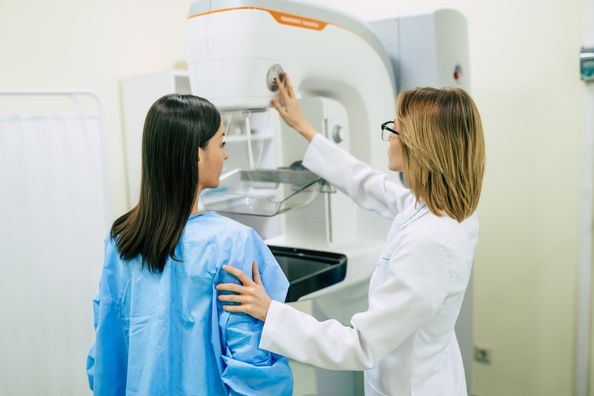
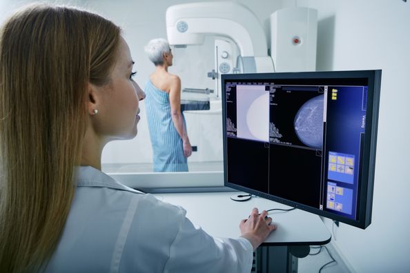
You’ve followed your physician’s recommendation and had your yearly screening mammogram performed, but then you get called back for additional imaging. Naturally your initial reaction causes your blood pressure to spike … this is a test that detects breast cancer after all. However, only 10% of women called back for more tests are found to have breast cancer according to the American Cancer Society. Learn more about call-backs and why it might not necessarily be cancer.
What is a “call-back” mammogram?
A call-back mammogram is a follow-up to an initial screening mammogram. Call-backs mean that more imaging or an ultrasound is needed to get a closer look at an area of concern. The point of the call-back is to provide a thorough examination of the abnormality and for precautionary measures. Getting called back can be common for women having their first mammogram due to lack of previous imaging for comparison or for pre-menopausal women that have dense breast tissue. An abnormal finding on a mammogram is not always cancer.
What else could the abnormality be?
An abnormal finding on a mammogram can be dense breast tissue, overlapping tissue, cyst, or non-cancerous mass. In other instances, if there is an abnormal or suspicious finding, the routine screening mammogram images might not provide enough detail for the radiologist to make an accurate diagnosis or because there are no previous mammograms to compare. With the new implementation of 3D mammography, radiologists are able to view your images better than ever before. This “slice-by-slice” imaging allows radiologists to see each section of the breast, which has been shown to decrease the number of call-back requests.
What will happen at the call-back appointment?
Generally a diagnostic mammogram is requested by the radiologist. Diagnostic mammograms include more images of the breast at various angles (depending on abnormality location) to concentrate on a specific area. A radiologist will be on-hand to advise the mammography technician, to ensure all the necessary images are captured to evaluate the abnormality.
Additionally, the radiologist may request a breast ultrasound and/or a breast MRI for a more specific view. This varies from patient to patient.
After your call-back exams are completed, the radiologist will speak to you regarding the results. If the radiologist cannot rule out cancer from the additional exam, generally a biopsy is recommended for further analysis.
What if I need a biopsy?
If a breast biopsy is deemed necessary, it still does not mean you have cancer. Statistics show most biopsy results are non-cancerous; however, a biopsy is the most accurate method to ensure the abnormality is not cancerous. During the biopsy, tissues around the abnormality are numbed and a small tissue sample is taken for microscopic examination. After the tissue samples are removed, a small metal clip is placed in the breast where the tissues were biopsied. The clips identify exactly what areas of the breast were biopsied. Additionally, for future mammograms, the clips show that a previous biopsy was performed in that area. Clips generally stay in forever unless surgically removed, are made of titanium and are about the size of a sesame seed.
Breast tissue samples are sent to a lab where a pathologist will examine them under a microscope and make a diagnosis. Biopsy results can take up to 14 days to complete. Once your physician receives the results, you will be contacted.
What happens if cancer is identified?
If cancer or pre-cancerous tissue is identified, you will be referred to a breast surgeon who will review your medical information and recommend an individualized treatment plan. You will also be contacted by a nurse navigator who will be able to assist with a myriad of care aspects including scheduling appointments to helping you understand your care options.
Call 630−545−7880 to schedule your mammogram today. To schedule an appointment with a breast surgeon, please call 630−790−1700.
Health Topics:


